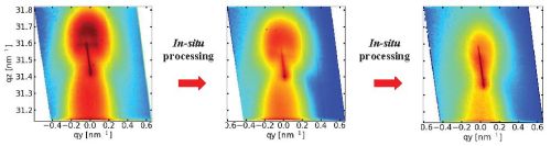
Time evolution (from left to right) of reciprocal space image of buried InGaAs/GaAs QDs around the 220 reflection during in-situ annealing.
In-situ investigations of self-organized nanostructures
The measurements are performed in UHV using RHEED, synchrotron radiation and optical methods.
Some literature overview:
T. Slobodskyy, A.V. Zozulya, R. Tholapi, L. Liefeith, M. Fester, M. Sprung, and W. Hansen, Versatile atomic force microscopy setup combined with micro-focused X-ray beam, Rev. Sci. Instrum. 86, 065104 (2015).
T. Slobodskyy, P. Schroth, D. Grigoriev, A.A. Minkevich, D.Z. Hu, D.M. Schaadt, and T. Baumbach. A portable molecular beam epitaxy system for in situ x-ray investigations at synchrotron beamlines. Rev. Sci. Instrum. 83, 105112 (2012).
P. Schroth, T. Slobodskyy, D. Grigoriev, A. Minkevich, M. Riotte, S. Lazarev, E. Fohtung, D. Z. Hu, D. M. Schaadt, T. Baumbach. Investigation of buried quantum dots using grazing incidence x-ray diffraction. Materials Science and Engineering: B 177, 721 (2012).
Suzuki, H. et al. Real-time observation of anisotropic strain relaxation by three-dimensional reciprocal space mapping during InGaAs/GaAs (001) growth. Appl. Phys. Lett. 97, 041906 (2010).
Fong, D.D., Lucas, C.A., Richard, M.-I. & Toney, M.F. X-Ray Probes for In Situ Studies of Interfaces. MRS Bulletin 35, 504-513 (2010).
Renaud, G., Lazzari, R. & Leroy, F. Probing surface and interface morphology with Grazing Incidence Small Angle X-Ray Scattering. Surf. Sci. Rep. 64, 255-380 (2009).
Richard, M.-I. et al. In situ x-ray scattering study on the evolution of Ge island morphology and relaxation for low growth rate: Advanced transition to superdomes. Phys. Rev. B 80, 045313-9 (2009).
Kaganer, V.M., Jenichen, B., Shayduk, R., Braun, W. & Riechert, H. Kinetic Optimum of Volmer-Weber Growth. Phys. Rev. Lett. 102, 016103-4 (2009).
Sasaki, T. et al. In situ Real-Time X-ray Reciprocal Space Mapping during InGaAs/GaAs Growth for Understanding Strain Relaxation Mechanisms. Appl. Phys. Express 2, 085501 (2009).
Chandril, S., Keenan, C., Myers, T.H. & Lederman, D. In situ thin film and multilayer structural characterization using x-ray fluorescence induced by reflection high energy electron diffraction. J. Appl. Phys. 106, 024308 (2009).
Image gallery:
GID maps at different exit angles of ring microstructures imaged by a microphocused X-ray beam.