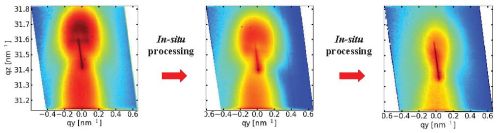
Time evolution (from left to right) of reciprocal space image of
buried InGaAs/GaAs QDs around the 220 reflection during in-situ annealing.
In-situ investigations of self-organized nanostructures
The measurements are performed in UHV using RHEED, synchrotron radiation and optical methods.
Some literature overview:
- T. Slobodskyy, A.V. Zozulya, R. Tholapi, L. Liefeith, M. Fester, M. Sprung, and W. Hansen, Versatile atomic force microscopy setup combined with micro-focused X-ray beam, Rev. Sci. Instrum. 86, 065104 (2015).
- T. Slobodskyy, P. Schroth, D. Grigoriev, A.A. Minkevich, D.Z. Hu, D.M. Schaadt, and T. Baumbach. A portable molecular beam epitaxy system for in situ x-ray investigations at synchrotron beamlines. Rev. Sci. Instrum. 83, 105112 (2012).
- P. Schroth, T. Slobodskyy, D. Grigoriev, A. Minkevich, M. Riotte, S. Lazarev, E. Fohtung, D. Z. Hu, D. M. Schaadt, T. Baumbach. Investigation of buried quantum dots using grazing incidence x-ray diffraction. Materials Science and Engineering: B 177, 721 (2012).
- Suzuki, H. et al. Real-time observation of anisotropic strain relaxation by three-dimensional reciprocal space mapping during InGaAs/GaAs (001) growth. Appl. Phys. Lett. 97, 041906 (2010).
- Fong, D.D., Lucas, C.A., Richard, M.-I. & Toney, M.F. X-Ray Probes for In Situ Studies of Interfaces. MRS Bulletin 35, 504-513 (2010).
- Renaud, G., Lazzari, R. & Leroy, F. Probing surface and interface morphology with Grazing Incidence Small Angle X-Ray Scattering. Surf. Sci. Rep. 64, 255-380 (2009).
- Richard, M.-I. et al. In situ x-ray scattering study on the evolution of Ge island morphology and relaxation for low growth rate: Advanced transition to superdomes. Phys. Rev. B 80, 045313-9 (2009).
- Kaganer, V.M., Jenichen, B., Shayduk, R., Braun, W. & Riechert, H. Kinetic Optimum of Volmer-Weber Growth. Phys. Rev. Lett. 102, 016103-4 (2009).
- Sasaki, T. et al. In situ Real-Time X-ray Reciprocal Space Mapping during InGaAs/GaAs Growth for Understanding Strain Relaxation Mechanisms. Appl. Phys. Express 2, 085501 (2009).
- Chandril, S., Keenan, C., Myers, T.H. & Lederman, D. In situ thin film and multilayer structural characterization using x-ray fluorescence induced by reflection high energy electron diffraction. J. Appl. Phys. 106, 024308 (2009).
Image gallery:
GID maps at different exit angles of ring microstructures imaged by a microphocused X-ray beam.